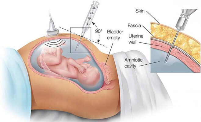
Amniocentesis
The main source of amniotic fluid is fetal urine. By analyzing a small sample of amniotic fluid, we can learn many things. For example, amniotic fluid studies can detect conditions such as Down syndrome, trisomy 18, trisomy 13 and other genetic abnormalities..
In most cases, you will be able to resume your normal activities the next day. The 20 milliliters of amniotic fluid that are removed account for no more than two to four percent of the total fluid in the uterus and your baby will replenish it within a day. If your blood type is Rh negative, we will recommend that you have a RhoGam injection before leaving the office.
What is amniocentesis?
- Amniocentesis is a procedure to remove small amount of fluid from around the fetus (amniotic fluid), for further testing. This fluid surrounds and protects your baby, also contain cells that originate from the fetus
How is amniocentesis done?
- You will usually receive genetic counseling before the procedure, to explain the genetic condition, and the procedure to be performed
- A very fine needle is passed into an appropriate pocket of amniotic fluid under continuous ultrasound guidance, to remove about 20 mls of the fluid.
- The procedure lasts about 1 minute and afterwards we would confirm that the fetal heart beat is normal.
- The fluid is mainly from fetal urine and it is expected to be reformed within a few hours.
- The procedure is performed after 16 weeks.
What should I expect after amniocentesis?
- There is about 1% risk of miscarriage within 5 days after the procedure, similar to the risk during CVS within the next five days.
- Amniocentesis performed before 16 weeks carries is a small risk of the baby developing club feet.
- Amniocentesis may be followed by mild abdominal discomfort, or menstrual-like pain which normally responds to paracetamol tablets and pregnancy continues
- If there is persistence, exacerbation of symptom or development of temperature, please seek medical advice.
- Generally, we advise bed rest for about 30 minutes after the procedure, avoidance of stressful physical activities for 3 days and sexual intercourse for at least 7 days.
When can I expect to get the results?
- The results for common genetic disorders would be ready within72 hours and 5 working days. The results for rare defects may take longer turn-around time.
Dating/viability scan
This is usually the first scan done after pregnancy is suspected. It is essentially to confirm the presence of a fetus(es) within the uterus and expected date of delivery. The scan can detect fetal heart activity from six weeks gestation by vaginal scan, and from eight to nine weeks by abdominal scan, while accurate dating can be obtained from measurements of the gestational sac and the ‘crown to rump’ length of the embryo.
Growth and Wellbeing Scan
This ultrasound scan is usually carried out between 32 -36 weeks of pregnancy. The scan is to confirm that the fetus is healthy and growing well. It is strongly advised that pregnancies that are complicated by disorders such as pre-eclampsia, abnormal fetal growth, diabetes mellitus, previous stillbirth, or any other adverse conditions during pregnancy should have the scan. Some obstetricians advise that this scan is offered to all women.
The aims of the scan are to:
- Measure the size of the fetal head, abdomen and thigh bone and calculation of an estimate of fetal weight
- Examine the movements of the fetus
- Evaluate the placental position and appearance
- Measure the amount of amniotic fluid
- Assess blood flow to the placenta and fetus by color Doppler ultrasound
