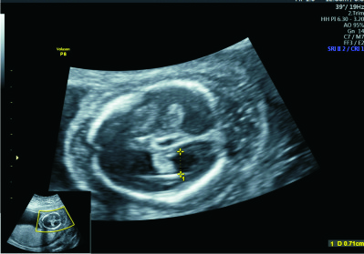
Anomaly Scan
This scan is also referred to as second trimester anomaly scan or level-II scan. It is a detailed scan done at 18-24 weeks during which we examine each part of the fetal body, to see if the baby has normal organs and also developing normally. The ultrasound machine, enables real time visualization of the fetal brain, face, spine, heart, stomach, bowel, kidneys and limbs, thus allowing for better interpretation of any abnormality. Special attention is devoted to the position of the placenta, and the amount of amniotic fluid.
Why do I need the Fetal Anomaly Scan?
Fetuses that have anomaly often do not reveal any background risk except in few cases. The only certain way to detect it therefore is through the anomaly scan. The anomaly scan is therefore recommended in all pregnancies, irrespective of the background status. Some of the other reasons for which the anomaly ultrasound scan, could be recommended, include suspicion of abnormality during a routine ultrasound exam, family history or investigation finding of a risk factor for anomaly such as maternal age, diabetes mellitus etc. Anomaly scan provides you a reasonable degree of assurance that your fetus is free from most major anomalies.
Is Anomaly scan safe for the fetus?
Yes anomaly scan does not put your fetus at any risk if it is done by the experts, working under standard safety guidelines. This is why we ensure that only trained hands conduct your scan.
What do I expect during the scan session
The scan takes about 40 -60 minutes. It is not painful, and does not require use of any sharp objects.
The sonographer will often times take you through the examination as it is being performed. Occasionally, the explanation is done at the end of the session.
Most of the fetal organs are seen during examination except few ones due to fetal position and activity.
The examination includes identifying fetal position, placental location and measurements of the head, abdomen and extremities. Additional procedure include color Doppler evaluation of blood flows and 3/4D Doppler.
What sort of problems can be found?
Examples of common problems seen during anomaly scan are shown in this section. Few organ abnormalities may not be well visualized at this time, due to reasons different from anomaly.
Can Down syndrome, and other chromosomal abnormalities be diagnosed at anomaly scan?
The anomaly scan is not for the diagnosis of Down syndrome or other chromosomal syndrome. However, it is useful to identify recognized soft ultrasound markers such as thick skin behind the neck (nuchal fold), absent nasal bone, mild fluid within the ventricles of the brain, aberrant subclavian artery in the neck, occasionally short arms or legs, white spots in the baby’s heart or abdomen, or choroid plexus cysts in the brain. The presence of these markers increases the chance that the fetus could be affected by Down syndrome. Please note that the presence of the markers is not a confirmatory test. Many normal babies also have these signs.
At the end of the scan
You pregnancy will be categorized into any of the following groups:
- Satisfactory outcome: which means that no major abnormality was diagnosed? You may however do the growth scan at 32 -36 weeks, for fetal monitoring
- Requires follow up scan: which means that there are still areas of the fetus that should be re-evaluated as pregnancy continues.
You will be adequately informed on your group and the steps to take.


